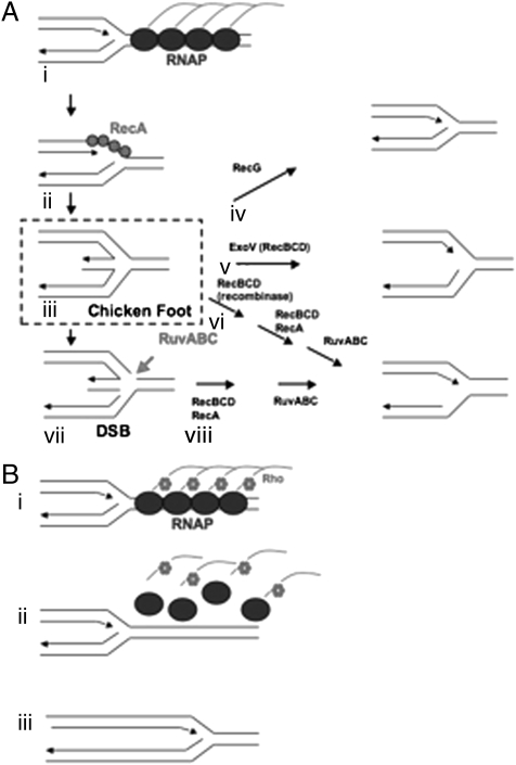
Fig. 1.
(A) Model of replication fork collapse and DSB formation. Adapted from Mahdi et al. (8) (i) Replication fork encounters TEC. The TEC arrest and form arrays that induce fork regression. (ii) RecA binds to exposed ssDNA and promotes recombination between leading and lagging strand forming “chicken foot” structures. (iii) “Chicken foot” structures are processed through multiple redundant pathways to allow reloading of replication forks. (iv) RecG separates leading and lagging strands and restores the fork. (v) RecBCD exonuclease activity degrades the hybrid, reforming the fork. (vi) RecBCD, RecA and RuvABC can restore the fork in a three step process without cleaving the Holliday junction. (vii) RuvABC cleaves the “chicken foot” structure across the Holliday junction. Failure to repair the “chicken foot” structure is proposed to lead to DSB formation. (viii) RecBCD, RecA and RuvABC restore replication forks in a two-step process. (B) Model of Rho-dependent release of TEC array. (i) Replication fork encounters an array of TEC promoter-proximal to an arrested TEC (7). (ii) Before the replication fork can regress, Rho factor removes the arrayed RNAP. (iii) Replication proceeds.

