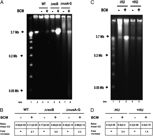
Fig. 4.
Pulsed-field gel analysis of BCM-induced DSBs. Cells were treated with 100 μg/mL BCM for 12 h where indicated. DNA fragments were separated on a 1% agarose gel in 0.5× TBE at 14 °C for 26.4 h at 6 V/cm (200 V) using a 120° included angle with a 2.8- to 26.3-s linear switch time ramp. Intact chromosomes remain in the well, and linearized DNA runs at 3.7 Mb. (A) PFGE. lane1: λ concateners from 0.05 to 1.0 Mb; lane 2: 3.7-Mb linearized E. coli chromosomes (MDS42 I-SceI); lane 3: MDS42; lane 4: BCM-treated MDS42; lane 5: MDS42ΔrecB; lane 6: BCM-treated MDS42ΔrecB; lane 7: MDS42 ΔnusA-G; lane 8: BCM-treated MDS42 ΔnusA-G. (B) Ratios of linearized to intact chromosomes. Ratio linear:cc = amount of linearized chromosomal DNA divided by amount of DNA retained in well. Fold-increase = fold-increase in linear DNA after BCM treatment. (C) PFGE of HU-treated Cells. MDS42 ΔrecB cells were prepared as above, except that cells were treated with 100 μg/mL BCM for 6 h, where indicated, in the presence or absence of 150 mM hydroxyurea (HU) to inhibit DNA replication. (D) Ratios of linearized to intact chromosomes. All values are the average of three or more independent experiments.

