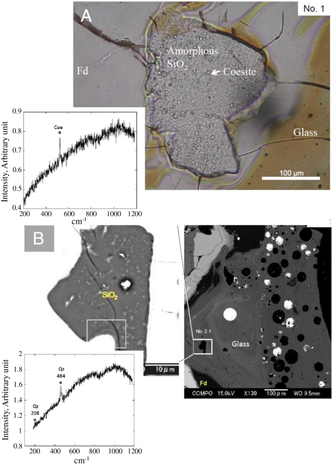
Fig. 2.
Amorphous silica grains No. 1 in the fragments, Asuka-881757-2 and 881757-3. (A) Amorphous silica grain, No. 1 contains many coesite inclusions with 1–10 μm in diameter. A Raman spectrum of an inclusion is shown in the Inset of this figure, showing the typical Raman peak of coesite at 522 cm-1. Fd, maskelynite with a feldspar composition; Glass, glass showing mixing of two melts with the pyroxene (brown color) and feldspar (gray) compositions. (B) a BSE image of amorphous silica grain No. 2. Inclusions of quartz were observed in this silica grain. SiO2, amorphous silica; A Raman spectrum of an inclusion is shown in the inset of this figure, showing the Raman peaks of quartz at 208 cm-1 and 464 cm-1. The major Raman peak observed at 464 cm-1 is larger than 456 cm-1 that is typical for the shock compressed quartz (8), suggesting that the quartz grains were formed by the back transformation from high-pressure phases.

