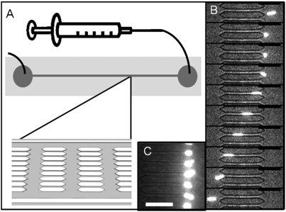
Fig. 2.
Microfluidic evaluation of RBCM deformability. (A) A schematic of the microfluidic devices, with flow driven by a syringe pump. (B) An image sequence showing a 1% cross-linked RBCM (6.0-μm diameter) deforming to pass through a 3 × 3.5 μm channel (25 ms between frames). (C) Ten percent cross-linked RBCMs stuck in the entrance of the 3-μm pores in a microfluidic device. Scale bars are 30 μm.

