Fig. 1.
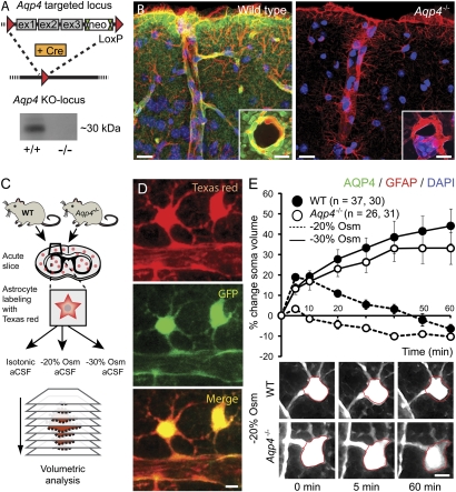
Aqp4 KO strategy and validation. (A) The targeted allele contained a flippase recognition target (FRT)-neomycin-FRT cassette after exon 3 and LoxP sites upstream and downstream of exons 1–3. Floxed mice were bred with Cre-expressing mice to produce mice with the Aqp4 KO allele. Western blot confirmed the absence of AQP4 in Aqp4−/− mice. (B) Immunofluorescence micrographs of mouse cortex probed with primary antibodies against AQP4 (green) and GFAP (red) with DAPI-labeled nuclei (blue) for orientation. The AQP4 immunofluorescence signal is absent in Aqp4−/− mice. Insets display perivascular AQP4 and GFAP labeling at higher magnification. (Scale bar: 25 μm; Inset, 5 μm.) (C) Experimental design for validating the effect of Aqp4 deletion on osmotically induced astrocyte swelling. Acute brain slices were prepared from WT and Aqp4−/− mouse pups. Slices were loaded with Texas red hydrazide and perfused with aCSF with normal (isotonic) or reduced osmolarity (−20% or −30% Osm). Sectional images were acquired for 3D volume analysis. (D) Two-photon imaging of Texas red hydrazide-loaded slices obtained from mice expressing GFP in astrocytes (Glt-1–EGFP BAC transgenic mice) confirmed that the dye was selectively taken up by astrocytes. (Scale bar: 5 μm.) (E) Exposure of acute cortical slices to 20% reduction in osmolarity (−20% Osm) induced more prominent swelling of astrocytic somata in WT (n = 37) than in Aqp4−/− mice (n = 26; P < 0.001 at 5 min, two-tailed Student t test). The initial swelling was followed by shrinkage reflecting regulatory volume decrease. More severe osmotic stress (−30% Osm) induced continuous swelling in both genotypes (n = 30 and 31). Error bars represent SEM. Lower shows representative images of astrocytes exposed to −20% Osm. The red ring marks the astrocyte soma circumference at baseline. (Scale bar: 5 μm.)

