Fig. 1.
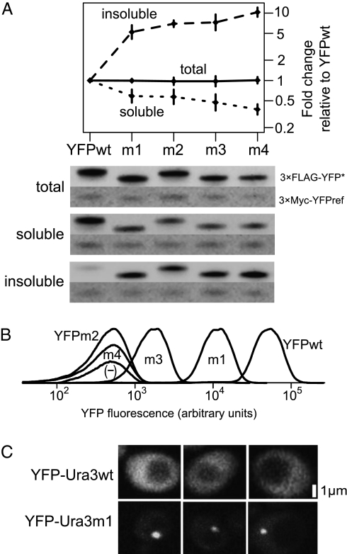
Protein variants display characteristic misfolding phenotypes. (A) Western blotting reveals partitioning of YFP variants into the insoluble fraction of a two-phase lysis. Blots of total (n = 2), soluble (n = 5), and insoluble (n = 4) cell fractions were probed from strains expressing 3×FLAG-YFP variants for 2 h, with a representative of each shown. Strains carried an rpn4 deletion (a regulator of the proteasome) and were treated with 100 μM of the proteasome inhibitor bortezomib during induction (43) to reduce misfolded protein degradation. A reference protein (PGAL1-3×Myc-YFPref) present in each strain was used to normalize galactose induction and loading between strains. Error bars show 95% confidence intervals about the mean reference-normalized fold change over YFPwt. (B) YFP variants fluoresce at different levels. Densities show single-cell variability across >10,000 cells, with arbitrary heights set to visually separate distributions. (C) YFP-Ura3wt dispersed throughout the cytosol, whereas YFP-Ura3m1 often localized in a single bright focus. Scale bar is estimated.

