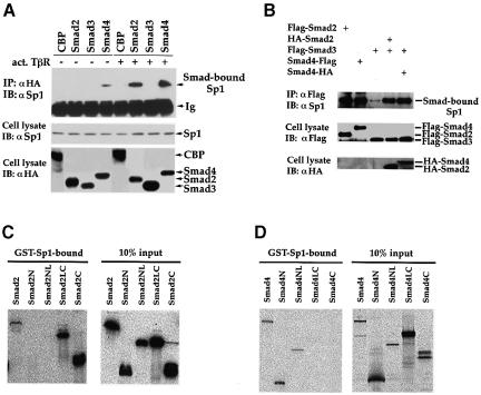
Fig. 4. Physical association of Smad2, Smad3 and Smad4 with Sp1. (A) TGF-β-dependent association of Smads and Sp1 in vivo. HaCaT cells were transfected with HA-tagged Smads or CBP, with (+) or without (–) an expression plasmid for activated TβRI. Cell lysates were immunoprecipitated (IP) with an anti-HA antibody, followed by immunoblotting (IB) with an anti-Sp1 antibody to detect Smad-bound Sp1 (upper panel). Cell lysates were also directly immunoblotted with anti-Sp1 or anti-HA antibodies to demonstrate expression of endogenous Sp1 (middle panel) or transfected Smads (lower panel). (B) Smad3 co-immunoprecipitates with endogenous Sp1 in the presence of Smad2 or Smad4. HaCaT cells were transfected with expression plasmids for Flag-tagged Smad3 and HA-tagged Smad2 or 4. Immunoprecipitation with anti-Flag antibody was followed by anti-Sp1 immunoblotting to detect Smad3-bound Sp1. Expression levels of Flag-tagged Smads (middle panel) or HA-tagged Smads (lower panel) were shown by anti-Flag or anti-HA immunoblotting. (C) Direct interaction of GST–Sp1(1–621) with 35S-labeled Smad2 and its segments. Smad2N, Smad2NL, Smad2LC and Smad2C cover the Smad2 regions of amino acids 2–183, 2–273, 181–467 and 270–467, respectively. (D) Direct interaction of GST–Sp1(1–621) with 35S-labeled Smad4 and its segments. Smad4N, Smad4NL, Smad4LC and Smad4C cover the Smad4 regions of amino acids 2–154, 2–300, 141–552 and 294–552, respectively.

An official website of the United States government
Here's how you know
Official websites use .gov
A
.gov website belongs to an official
government organization in the United States.
Secure .gov websites use HTTPS
A lock (
) or https:// means you've safely
connected to the .gov website. Share sensitive
information only on official, secure websites.
