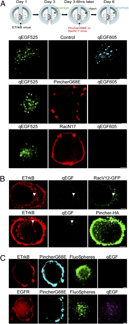
Fig. 2.
ETrkB, but not EGFR, retrograde transport requires both Pincher and Rac functions. (A) Chamber-cultured SCG neurons were infected with ETrkB adenovirus, the distal axons treated with qEGF525 for 6 h and neurons infected with either PincherG68E-HA or RacN17-T7 adenoviruses for 3 d, and distal axons treated for 2 h with qEGF605 (see schematic). Immunocytochemistry: qEGF525 (green), qEGF605 (cyan), PincherG68E (anti-HA, red, Top and Middle), and RacN17 (anti-T7, red, Bottom). (B) Distal axons of chamber-cultured SCGs coinfected with adenoviruses expressing ETrkB and either RacV12-GFP (Upper) or Pincher-HA (Lower) were treated with qEGF for 2 h. Immunocytochemistry: ETrkB (anti-EGFR, red), qEGF (cyan), RacV12-GFP (GFP, green, Upper) and Pincher-HA (anti-Pincher, green, lower). Arrowheads point to receptor/qEGF complexes. (C) Distal axons of chamber-cultured SCGs coinfected with adenoviruses expressing PincherG68E-HA or either ETrkB (Upper) or EGFR (Lower) were treated for 48 h with fluoSpheres and then with qEGF for 2 h. ETrkB (anti-EGFR, red, Upper), EGFR (anti-EGFR, red, Lower), PincherG68E (anti-Pincher, cyan), fluoSpheres (green), and qEGF (purple). (Scale bars, 2 μm.)

