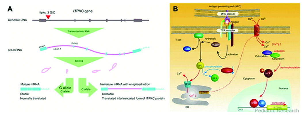
Figure 2.
Functional significance of itpkc_3 on ITPKC mRNA and Ca2+/NFAT pathway. (A) Effect of itpkc_3 C allele on splicing of ITPKC pre mRNA. The C allele of itpkc_3 reduces splicing efficiency of IPTKC premRNA. mRNAs harboring unspliced intron 1 cannot be translated properly and will be degraded early by nonsense-mediated decay mechanism. (B) Proposed role of ITPKC as a negative regulator of Ca2+/NFAT pathway. When the T-cell receptor (TCR) is bound by antigen/MHC complex on antigen presenting cells (APCs), adaptor molecules and kinases are recruited and phospholipse C-γ1 (PLC-γ1) is activated by phosphorylation of its tyrosine residue. IP3 and diacylglycerol (DAG), another second messenger molecule, are generated by hydrolysis of phosphatidylinositol 3,4-bisphosphate (PIP2) by activated PLC-γ1. IP3 binds to its receptor expressed on endoplasmic reticulum (ER) membrane and causes the release of Ca2+ into the cytoplasm. Then depletion of Ca2+ store in ER evokes a process termed as store operated Ca2+ entry in which extracellular Ca2+ enters through calcium release-activated Ca2+ channels on the plasma membrane. Recent advances in research identified the role of stromal interaction molecule (STIM) as a sensor of Ca2+ in ER and ORAI as a calcium release-activated Ca2+ channel. Cytoplasmic Ca2+ binds calmodulin, which in turn activates calcineurin, a calmodulin-dependent phosphatase. Activated calcineurin dephosphorylate NFAT in the cytoplasm and lead nuclear translocation of NFAT. NFAT in the nucleus drives transcription of genes important in T cell activation as a homodimer or heterodimer with other transcription factors. AP1 is one of the transcription partners of NFAT, which is activated by a signal from TCR mediated by DAG (72–74). Reactions and amounts of molecules increased by the effect of itpkc_3 C alleles were represented by red characters and arrows and those reduced by blue, respectively. [Ca2+]i: intracellular free Ca2+ concentration. Reprinted with permission from Onouchi Y. Molecular Genetics of Kawasaki Disease. Pediatric Research 65(5 Part 2):46R–54R, 2009.

