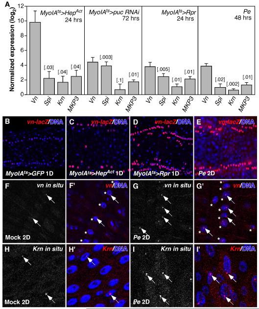
Figure 1. Drosophila EGFR ligands are induced in the regenerating adult midgut.
A. RT-qPCR quantification of Drosophila EGFR ligands (vn, spi and Krn) and MKP3 (MAP kinase phosphatase-3) mRNA expression in the regenerating midgut. The midgut was induced to regenerate by activating the JNK pathway in the ECs (MyoIAts>HepAct, 24hrs or puc RNAi, 72hrs), or inducing EC apoptosis (MyoIAts>Rpr, 24hrs), or Pe infection (48hrs). STDEV and P-value (t-test) were shown. B-E. Expression of vn-lacZ reporter in control (B) or regenerating posterior midguts (C-E). 2 of the 4 rows of circular visceral muscle cells (VM) were shown. F, G. vn fluorescent in situ hybridization. The strongest vn signals were in the nucleus (arrows) of VMs (asterisks), most likely the loci of Vn transcription. H, I. Krn fluorescent in situ hybridization. The strongest Krn signals were in the nucleus of ECs (arrows). In mock-infected control midguts, vn and Krn were expressed at low levels in the VM and ECs respectively (F, H).

