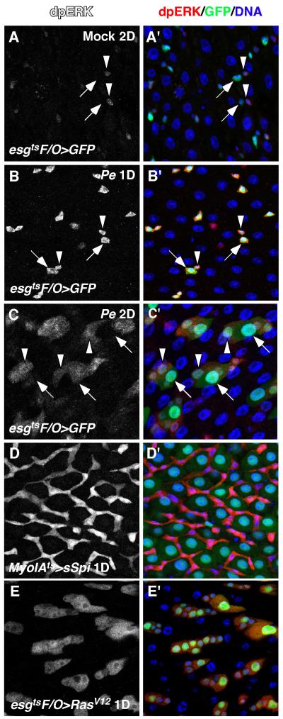
Figure 2. MAPK is activated in the regenerating midgut.
The activity of Drosophila MAPK was assayed by anti-dpERK staining. A, A’. MAPK activity in the mock-infected control midgut. B, B’. MAPK activity after infecting with Pe for 1 day. ISCs and EBs were marked by esgGal4-driven GFP expression and indicated by arrowheads and arrows respectively (A, B). C, C’. MAPK activity after infecting with Pe for 2 days. Differentiating ECs (pre-ECs, medium nucleus) and newly formed mature ECs (large nucleus) were indicated by arrowheads and arrows respectively. D, D’. MAPK activation induced by ectopic expression of sSpi (MyoIAts>sSpi). E, E’. Cell autonomous MAPK activation induced by activated Ras (esgtsF/O>RasV12).

