
Figure 1. Increased EC and pericyte density in retinas from mature bim-/- mice.
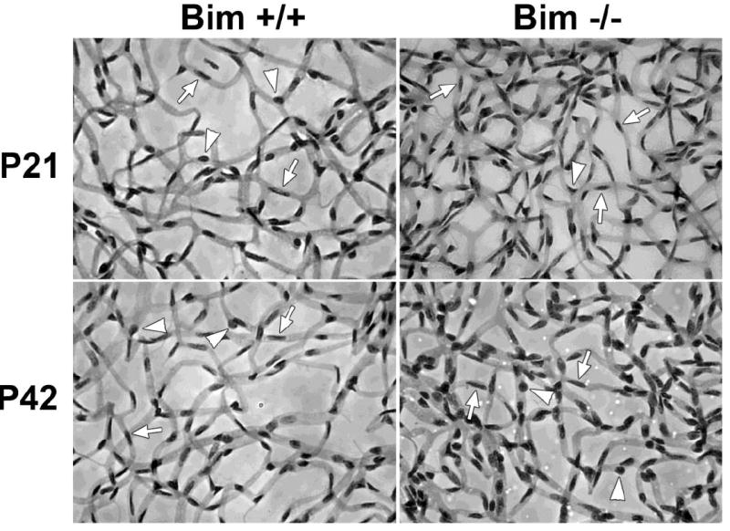
Retinas from P21 and P42 bim +/+ and bim -/- mice were prepared by trypsin digest and HE/PAS staining (Wang et al., 2005; Wang et al., 2003). EC and pericytes were then quantified per 100 μm2. Please note that retinas from bim -/- mice demonstrate increased numbers of EC (arrows) and pericytes (arrow heads). Experiments were repeated with eyes from 5 mice with similar results. The quantitative assessment of the data is summarized in Tables 1-3.