
Figure 2. The spreading of superficial retinal blood vessel layer appears similar in P7 bim +/+ and bim -/- mice.
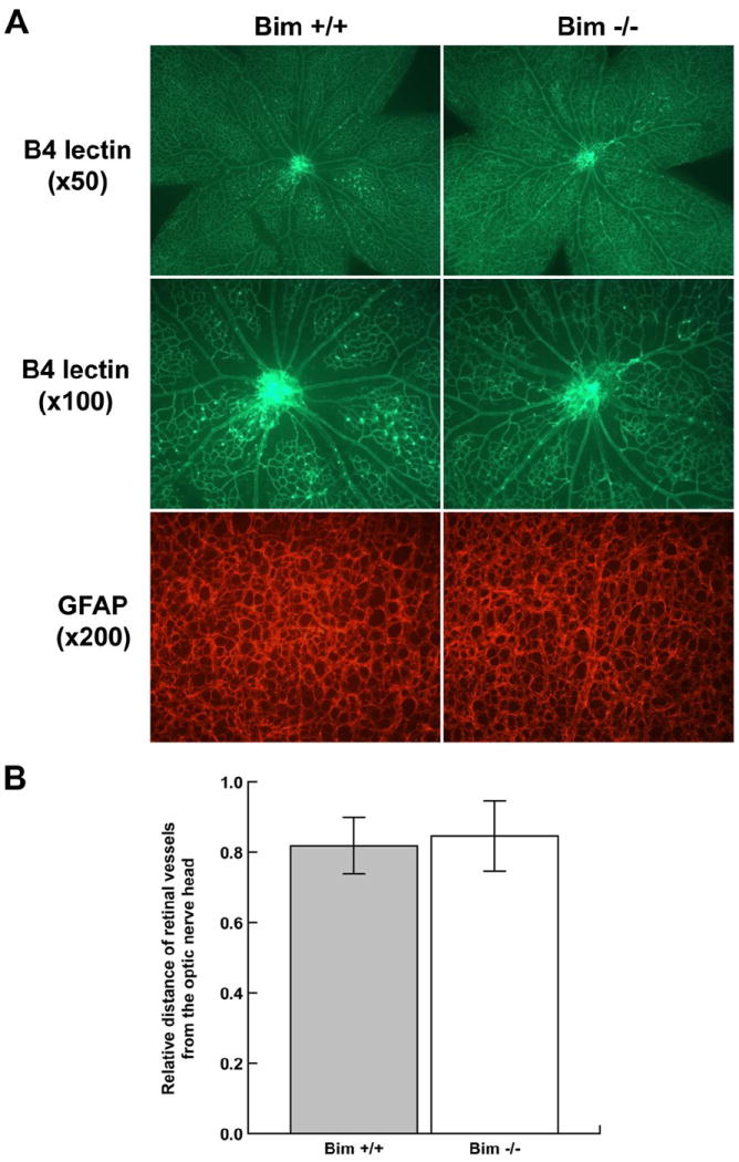
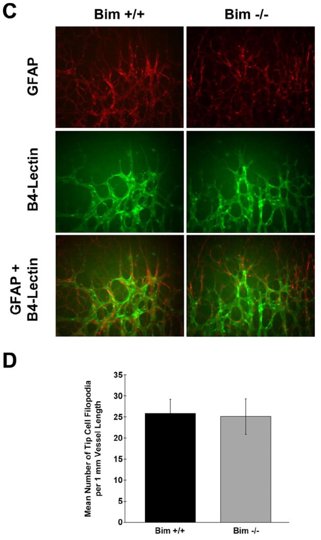
In Panel A, assessment of retinal vasculature was performed by wholemount staining with B4 lectin. These experiments were repeated at least three times with eyes from three different mice. Astrocyte density and organization was assessed by GFAP staining. Panel B determines the relative distance the retinal vessels have spread from the optic nerve head. In Panel C, retinas from P5 mice were wholemount stained with B4-lectin and anti-GFAP as noted. Panel D summarizes the mean number of tip cell filopodia per 1mm vessel length obtained from data presented in Panel C. Please note that the retinal vasculature appears similar in terms of appearance, organization and tip cell density in bim +/+ and bim -/- mice at this time point. Experiments were repeated with eyes from 5 mice with similar results.