
Figure 4. Active Cdc42 improves capillary morphogenesis by inhibiting GSK-3β.
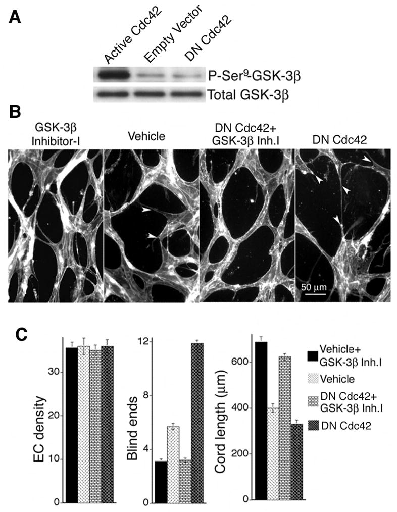
(A) Active L28Cdc42 inhibits GSK-3β in MVECs, as indicated by Ser9 phosphorylation. By scanning densitometry, active L28Cdc42 increased phosphorylation of GSK-3β Ser9 > 10-fold relative to controls in three separate experiments. Conversely, DN Cdc42 reduced GSK-3β Ser9 phosphorylation ~ 50%. (B) GSK-3β inhibitor-I (3μM) was added to control and DN N17Cdc42 MVECs “sandwiched” between two layers of collagen-I in the presence of VEGF. Note absence of blind ends (arrows) in the presence of GSK-3β inhibitor-I. (C) Quantification of cord parameters; n ≥ 22 for all groups. GSK-3β inhibitor-I significantly improved formation of capillary cords, as measured by increased cord length and reduction in cord blind ends (p < 0.001 for all paired comparisons between GSK-3β inhibitor-I and the corresponding control).