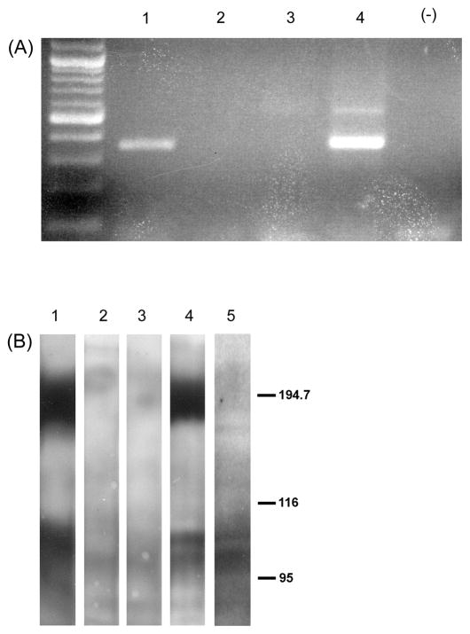
Figure 2.
A. rtPCR analysis of transcripts from non-transfected C6 cells (lanes 2 and 3) and two C6 cells stably transfected with mGluR2 (lanes 1 and 4). Major DNA bands in lanes 1 and 4 are in the size range predicted for mGluR2 rtPCR products obtained via the primers used in this study.
B. Western blot analysis of protein from non-transfected C6 cells (lanes 2 and 3), two C6 cell lines stably transfected with mGluR2 (lanes 1 and 4) and one C6 cell line stably transfected with mGluR3 (lane 5). Blots were reacted with mGluR2/3 antibody (Chemicon). Under these experimental conditions, mGluR2 receptors migrate more as dimers (~200 kD) than monomers (~100 kD) while mGluR3 tends to migrate primarily as the monomer (~100 kD).

