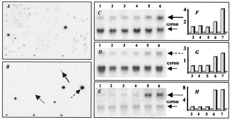
Figure 3.
Expression profiles for genes isolated from RV-infected mouse brain by SH. Hybridization of 32P-labeled cDNA probes from normal brain with 6 day infected brain cDNA subtracted (A) and from RV-infected brain at day 6 p.i. with normal brain cDNA subtracted (B) to Atlas cDNA mouse 1.2 Expression Array. Northern blot analysis of genes identified by SH (C–E). Total RNAs prepared from uninfected brain (lanes 1) or infected for 3 days (lane 2), 4 days (lane 3), 5 days (lane 4), 6 days (lane 5), and 7 days (lane 6) were hybridized with 32P-labeled probes for semaphorin G (solid line in C and B); laminin receptor 1 (dashed line in D and B); neuroleukin (dotted line in E and B). Expression values for each gene (F–H) were determined as described in the legend to Fig. 1.

