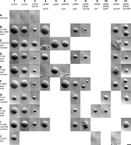
FIG. 2.
Photomicrographs of F. johnsoniae colonies. Colonies were grown for 48 h at 25°C on PY2 agar medium containing the appropriate antibiotics. Photomicrographs were taken with a Photometrics Cool-SNAPcf2 camera mounted on an Olympus IMT-2 phase-contrast microscope. Bar (at lower right; applies to all panels), 1 mm. Rows (A to J) indicate different strains of F. johnsoniae, and columns (1 to 12) indicate different plasmids introduced into these strains. Rows: A, wild-type F. johnsoniae UW101; B, fjoh_0983 insertion mutant CJ1708; C, sprC mutant UW102-91; D, sprC HimarEm2 insertion mutant FJ105; E, sprD HimarEm2 insertion mutant FJ162; F, sprD insertion mutant CJ1695; G, sprB HimarEm2 insertion mutant FJ114; H, sprB HimarEm2 insertion mutant FJ156; I, sprCDB deletion mutant CJ1584; J, sprF insertion mutant CJ1814. Columns: 1, control vector pCP23; 2, control vector pCP29; 3, control vectors pCP23 and pCP29; 4, pSN80 expressing sprC and sprD; 5, pSN81 carrying sprC and sprD in the opposite orientation as pSN80; 6, pMM339 expressing sprD; 7, pSN60 expressing sprB; 8, pSN80 (sprC sprD) and pSN60 (sprB); 9, pRR48 expressing sprF; 10, pRR48 (sprF) and pSN60 (sprB); 11, pRR49 expressing sprC, sprD, and sprF; 12, pRR49 (sprC sprD sprF) and pSN60 (sprB).

