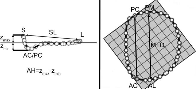
Fig 2.
Annular modeling. (A) Intercommissural and (B) transvalvular views of a real-time three-dimensional echocardiography derived mitral annular model are depicted. The least squares plane has been superimposed on the annulus in each view. (A) The manner in which the annular height (AH) and septolateral diameter (SL) are determined is illustrated. (B) The manner in which the intercommissural width (CW) and the mitral transverse diameter (MTD) are determined is illustrated. (AC = anterior commissure; AL = anterolateral annulus; L = lateral annulus; PC = posterior commissure; PM = posteromedial; S = septum; Zmax = maximum annular height corresponding to septum; Zmin = minimum annular height.)

