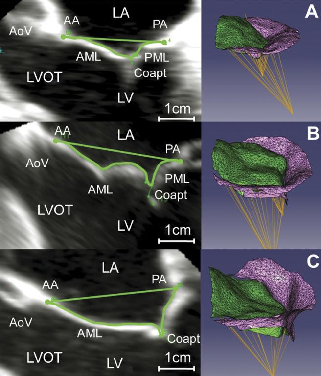
Fig 3.
Three-dimensional valve renderings. Mitral leaflet tethering for representative valves: (left) two-dimensional echocardiographic image with tenting area delineated; (right) three-dimensional rendering in an oblique view. (A) Normal valve, (B) ischemic mitral regurgitation (IMR) valve with limited tethering, and (C) IMR valve with severe leaflet tethering. (AA = anterior annulus; AML = anterior mitral leaflet; AoV = aortic valve; Coapt = coaptation; LA = left atrium; LV = left ventricle; LVOT = left ventricular outflow tract; PA = posterior annulus; PML = posterior mitral leaflet.)

