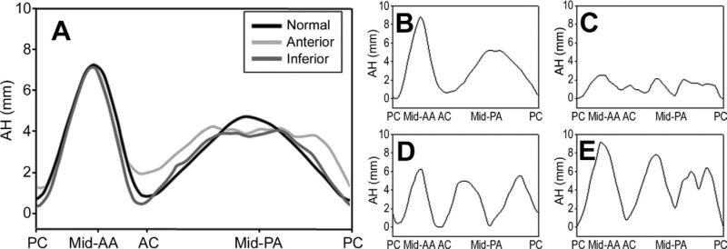
Fig 4.
Regional annular shape, presented as regional annular height (AH) plotted as a function of rotational position, averaged for (A) cohorts and for (B–E) selected ischemic mitral regurgitation patients. All patients’ commissures were aligned in such a way that the posterior commissure (PC) was located at 0% and the anterior commissure (AC) at 33.3% of the rotational position along the annulus. Note the increased variability of AH in the posterior annular (PA) region. Also note the extremely variable annular shape, varying from normal (B) to flat (C), mid-posterior restriction (D), and bimodal posterior restriction (E). (AA = anterior annulus; PC = posterior commissure; black line = normal; dark gray line = inferior; light gray line = anterior.)

