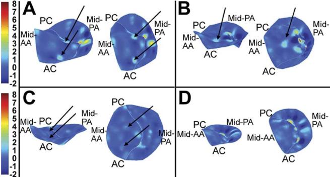
Fig 6.
Three-dimensional renderings of leaflet curvature. Color contouring is determined by three-dimensional leaflet Gaussian curvature (K). Negative K values indicate saddle-shaped (hyperbolic) local surface curvature; positive K values indicate elliptic local surface curvature but may be concave toward the left ventricle or left atrium. Valves are rendered as if the viewer were looking down from the left atrium. The left-hand image in each panel is oriented to allow appreciation of the three-dimensional directionality of positive Gaussian curvature (see text); the right-hand image is a two-dimensional planar projection of the leaflets. (A) A valve with two areas of intensive positive curvature convex toward the left ventricle; this was the most common configuration in the ischemic mitral regurgitation group. (B) A unifocal area of intense positive curvature is demonstrated. (C) A lesser degree of positive curvature foci is illustrated. (D) A healthy person with normal leaflet redundancy is shown. (AA = anterior annulus; AC = anterior commissure; PA = posterior annulus; PC = posterior commissure.)

