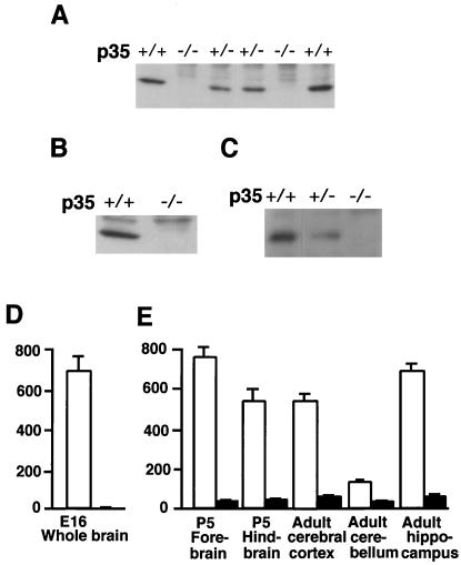
Figure 2.
p35−/− mice retain residual Cdk5 kinase activity. (A–C) Western blot analysis of p35 protein of the brain homogenate (A and B) and immunoprecipitate with anti-Cdk5 antibody (C) from the indicated genotype at postnatal day (P) 2 by using p35 antibodies that recognize the carboxyl terminus of p35 protein (A and C) and the N terminus of p35 protein (B). No p35 protein was detected in p35−/− brain, and a reduced amount of p35 protein was detected in the p35+/− brain (A and C). (D and E) Cdk5 kinase activity in brain homogenates from Cdk5+/+ (white bar) and Cdk5−/− (D) and p35+/+ (white bar) and p35−/− (black bar) homogenates (E). The diagram represents mean ± standard deviation (n = 3) of Cdk5 activity. Cdk5 kinase activity is expressed as pmol of phosphate incorporated per hr per mg protein. Approximately 10 and 20% residual Cdk5 kinase activity is detected in the cerebral cortex and cerebellum of p35−/− brain, respectively, but none in the Cdk5−/− brain.

