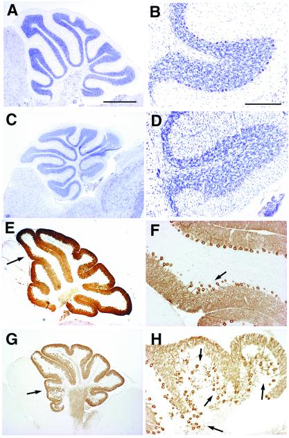
Figure 4.
Migration defects of cerebellar cortical neurons in p35−/− mice are accentuated by Dab1 heterozygosity. Comparative histology of cerebella of Dab1yot/+ (A and B), p35−/− (E and F), and p35−/−Dab1yot/+ (C, D, G, and H) at P21 in the parasagittal sections stained with toluidine blue (A–D) and with anti-IP3R antibody (E–H). B, D, F, and H are higher magnifications of A, C, E, and G, respectively. Arrows in E and G indicate magnified areas in F and H, respectively. Subtle disturbances of the alignment of Purkinje cells are observed in p35−/− (arrows in F). Extensive migration defects of Purkinje cells and granule cells are observed in p35−/−Dab1yot/+ compared with p35−/− cerebellum (D, G, and H). (Bar in A, 1 mm. A, C, E, and G are identical magnification. Bar in B, 200 μm.)

