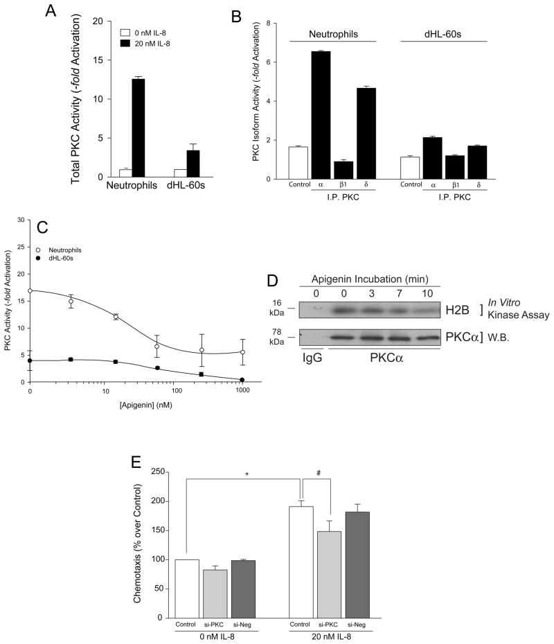
Fig. 4. IL-8 also activates PKC, albeit at a lower extent that it does JAK3.
(A) IL-8 stimulates total PKC activity of human neutrophils and differentiated HL-60 cells. Cells were incubated without or with 20 nM IL-8, lysates prepared and immunoprecipitated and PKC kinase reactions performed as in Materials and Methods. (B) Neutrophil and dHL-60 cell lysates were immunoprecipitated with anti-myc-agarose (negative control) or with α-PKCα-, α-PKCβ1- or α-PKCδ-agarose and used in the PKC kinase assay. (C) Effect of apigenin on PKC activity of neutrophils (open circles) and dHL-60 cells (filled circles). (D) dHL-60 cell lysates were immunoprecipitated with anti-PKCα antibodies or IgG control and subjected to in vitro kinase assay using H2B as substrate in the presence of [γ-32P] ATP. Kinase reactions were resolved by SDS-PAGE, western-blot transferred and products visualized using autoradiography. Autoradiograph of increasing time of incubation with apigenin on PKCα phosphorylation of H2B (top panel). The same membrane was immunoblotted with anti-PKCα antibody to ensure equal protein loading control (bottom panel). (E) Effect of silencing PKC with dsRNA for fours days on dHL-60 cell chemotaxis. Si-Control is a scrambled dsRNA.

