


Figure 2.
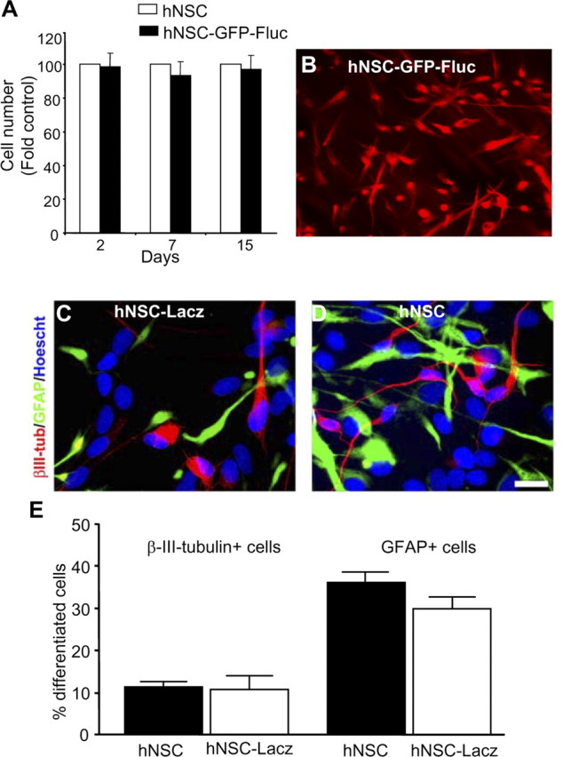
Characterization of transduced hNSCs. A, B, hNSCs were transduced with LV-GFP-Fluc, and growth of transduced and nontransduced cells in culture was compared over 15 d (A); immunohistochemistry was performed with anti-nestin antibody and detected with Alexa-540-conjugated secondary antibody (B). C, D, hNSCs or LV-Lacz transduced hNSCs were differentiated for 7 d, and immunocytochemistry was performed with βIII-tubulin and GFAP antibodies and detected with CY3- or CY5-conjugated antibody. Cell nuclei were counterstained with Hoescht 33258. E, Percentage of βIII-tubulin+ cells and GFAP+ cells. Data represent means ± SEM (n = 4). Magnification: B, 20×; C, D, 40×.