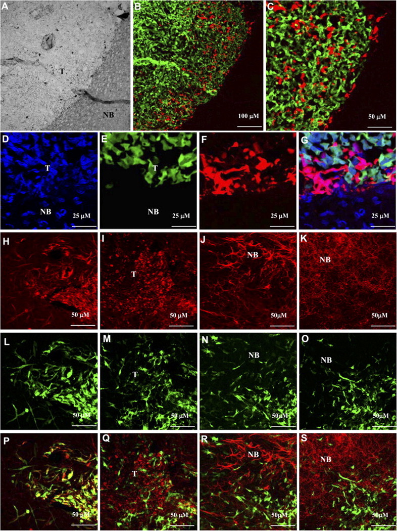
Figure 7.

hNSCs are present specifically in the gliomas in an undifferentiated state. Mice implanted with Gli36-GFP-Rluc glioma cells stereotactically into the right frontal lobe were implanted with Fluc-DsRed2 hNSCs 2 d later. Mice were imaged by intravital microscopy (see Fig. 6) and killed on day 10 after hNSC implantation. Mice brains were sectioned, and confocal microscopy was performed. A, Light image showing the normal brain (NB) and the tumor (T). B, Fluorescent image of A showing hNSCs (red) infiltrating the tumor (green); 10× magnification. C, Higher magnification (40×) image of B. D, Brain sections were stained with 1 μm TO-PRO-3 (nucleic acid staining) and confocal microscopy was performed. D, TO-PRO staining (blue). E, GFP-positive glioma cells. F, DsRed2 hNSCs. G, Merge of D–F. H–S, Immunohistochemistry on brain sections from Gli36-glioma-bearing mice implanted with hNSCs expressing GFP-Fluc, 10 d after implantation. Representative images of brain sections immunostained for nestin (H, L, P), Ki67 (I, M, Q), GFAP (J, N, R), and MAP-2 (K, O, S) are shown. Green, GFP expression; red, Ki67, GFAP, or MAP-2 expression; yellow, coexpression of GFP and nestin.
