
Figure 2. MI and ischemia/reperfusion (I/R) causes more cell death in β2a hearts.
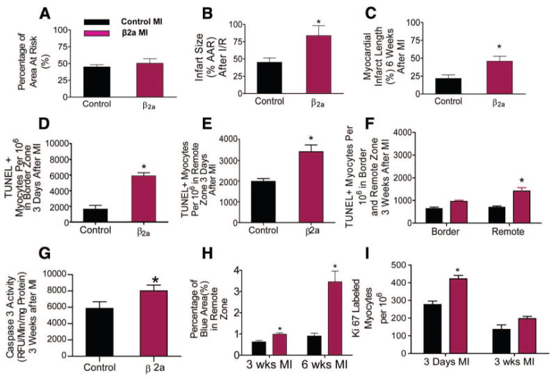
A, AAR was not different in control (n=15) and β2a hearts (n=6). B, Infarct size (% of AAR) in hearts with 30 minutes of ischemia followed by 24 hours of reperfusion (I/R) was greater in β2a (n=6) than in control hearts (n=8). C, Infarct length after permanent occlusion was greater in β2a (n=6) than in controls (n=7) and. D through F, Myocytes per 106 undergoing apoptosis in the border and remote zones of control (n=3) and β2a hearts (n=4). G, Caspase 3 activity in remote zone tissues (control, n=9; β2a, n=10). H, Fibrotic area (blue) in remote zones of trichrome-stained cardiac histological sections from control (n=3) and β2a hearts (n=3). I, Ki67+ myocytes per 106 in control (n=3) and β2a hearts (n=4) after MI. *P<0.05; **P<0.01.