
Fig. 6.
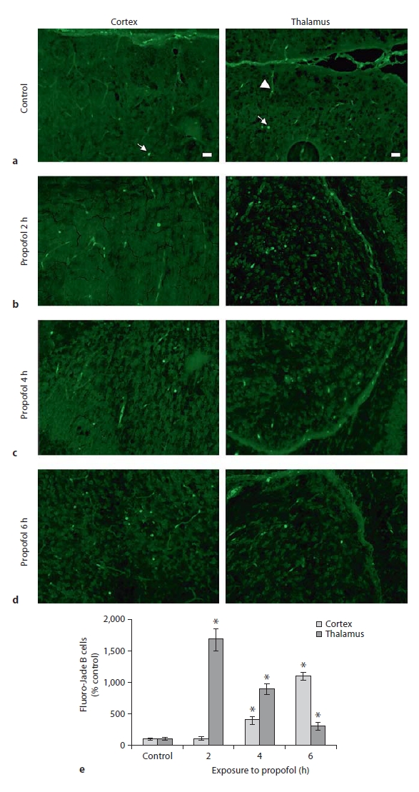
Fluoro-Jade B staining of P7 rat brains immediately (0 h) after termination of the 2-, 4- and 6-hour exposures to propofol. An increase in the number of Fluoro- Jade B-positive cells was apparent in the retrosplenial cortex following 4 and 6 h of exposure (c, d, left panel), and in the laterodorsal thalamic nucleus following 2-, 4- and 6-hour exposure (b, c, d, respectively, right panel). Quantitative analysis of Fluoro-Jade B staining at the retrosplenial cortex and the laterodorsal thalamic nucleus is shown (e). For each condition, 3 animals were randomly assigned. Data are presented as means ± SEM. A probability of * 0 < p.05 was considered significant. Degenerating neurons are marked with arrows, arrowheads point to blood vessels. Scale bar = 20 μm.