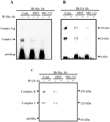
Figure 6. Androgen effects on the ubiquitination of p66Shc proteins.
LNCaP C-33 cells were plated in regular medium for 72 h, steroid starved for 48 h and then treated with or without DHT (10 nM) and MG 132 (0.5 µM) as a positive control for 24 h. Cell were harvested in lysis buffer. Total cellular lysates (300 µg proteins) were reacted with (A & B) anti-Shc or (C) anti-Ubiqutin Ab (3 µg each), and followed by Protein A–Sepharose beads. The immune complexes were analyzed by immunoblotting with (A) anti-Shc Ab, (B) anti-ubiquitin Ab and (C) anti-Shc Ab. The positions of p66Shc and p66Shc/Ub-protein complex are indicated by arrow-heads. The figure is a representative of four sets of independent experiments.

