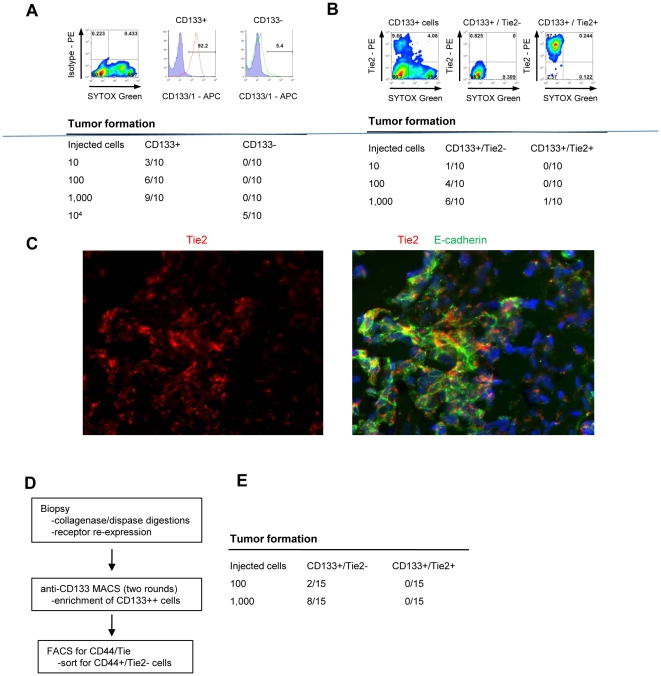
Figure 5. Tumor forming ability of CD133+ subfractions.
A) Validation of CD133+ cell fractions obtained by column isolation. Purity of injected cell fractions is shown. Cells used for transplantation were sorted by FACS and non-viable cells were excluded using SYTOX Green. Tumor-forming cells are highly enriched in the CD133+ cell fraction. Shown are studies with CD133+ vs CD133− cells from eight pooled ovc316-X xenografts. (N = 10 animals per group). Tumor formation was evaluated 4 months after inoculation. TIC CD133+: 1/191, TIC CD133−: 1/16725. Chi-square = 8.25, p<0.01. B) Column-isolated CD133+ cells were further subdivided into Tie2+ and Tie2− fractions by FACS. Cells used for transplantation were sorted by FACS and non-viable cells were excluded using SYTOX Green. Tumor-forming abilities are more pronounced in CD133+/Tie2− cells. Shown are data from transplantation of CD133+/Tie2+ vs CD133+/Tie− cells from ovc316-XC (N = 10). TIC CD133+/Tie2+: 1/10593, TIC CD133+/Tie2−: 1/650. Chi-square: 6.74, p<0.01. C) Immunofluorescence analysis of tumors that developed after transplantation of CD133+/Tie− cells. D and E) Analysis of fractions derived from patient biopsies. D) Schematic of cell isolation. E) Tumor formation after transplantation of CD133+/Tie2− and CD133+/Tie2+ cells derived from ovc100506-biopsy. Tumor formation was evaluated 3 months after inoculation. TIC CD133+/Tie2−: 1185, TIC CD133+/Tie2+: n.a.

