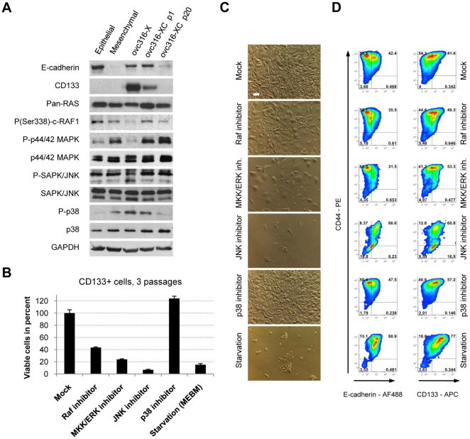
Figure 8. Impact of MAP kinases on proliferation and differentiation in CD133+ derived cells.
A) Western blot analysis of E-cadherin, CD133 and the MAPK pathway in ovc316-XC tumor xenografts, low passage (p1), high passage (p20), and in clonal epithelial and mesenchymal cultures. Xenograft tumors contain high amounts of E-cadherin and CD133, which decrease after culturing. Concomitantly with a loss of these markers occurs a switch from phophorylated p38 to phophorylated ERK44/42. B) Impact of MAPK pathway inhibitors on proliferation. Cells (p7) were passaged in the presence of inhibitors for 21 days. C) Bright field analysis of ovc316-XC cells cultured in the presence of indicated inhibitors after 21 days. The scale bar is 40µm. D) Flow cytometry analysis of ovc316-XC cells cultured in the presence of indicated inhibitors after 21 days (N = 3).

