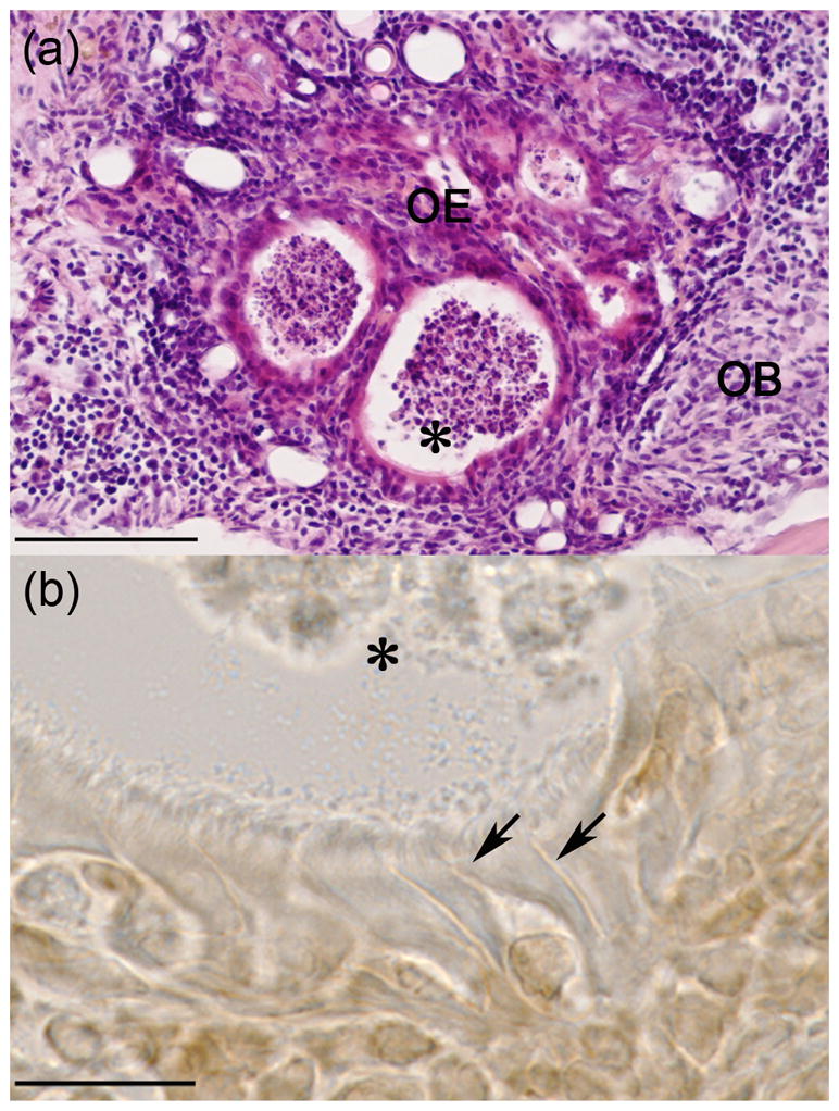
Figure 1.

Histological sections of OE graft in OB of a mouse at 30 day survival time point. A. H&E stained section of OE graft tissue showing formation of circular vesicles lined with epithelial cells. B. High magnification of epithelial cells stained for the Olfactory Marker Protein (OMP) confirming the presence of olfactory sensory neurons with characteristic vessel shape, apical dendrites (arrows) and olfactory cilia. Astric (*) marks a common reference point in images A and B. Scale bars: A, 100um; B, 15um.
