Figure 1.
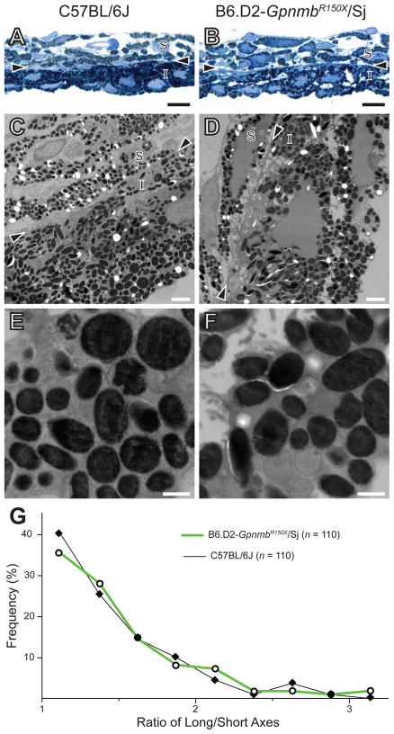
Lack of Gpnmb-mutant phenotype detectable by TEM in melanosomes of the iris. (A,B) Light microscopy (scale bar = 10 μm) and (C,D) TEM (scale bar = 2 μm) images showing similarities in the appearance of melanosomes from C57BL/6J (left column) and B6.D2-GpnmbR150X/Sj (right column) irides. (E,F) Higher magnification TEM of iris pigment epithelium (scale bar = 0.5 μm). Arrowheads, border region; S, iris stroma, I, iris pigment epithelium. (G) Shape distribution plot showing that in situ melanosomes of the iris from C57BL/6J and B6.D2-GpnmbR150X/Sj mice share a similar shape.

