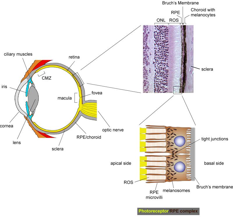
Figure 1.
Anatomy of the camera-style mammalian eye and photoreceptor/retinal pigment epithelium (RPE) complex. Left: schematic cross-section through the human eye showing its main structural components that allow focusing of the light onto the photoreceptor layer of the retina. Top: Histologic section of an adult mouse eye showing photoreceptor nuclei in the outer nuclear layer (ONL), rod outer segments (ROS), RPE, Bruch’s membrane, and the choroid with its neural crest-derived melanocytes. Bottom: Diagram showing two RPE cells with their microvilli contacting the rod outer segments. The intimate anatomic relationship of RPE cells with photoreceptors and Bruch’s membrane allow the cells to have multiple roles as support cells for the retina (see text). The cells also carry a dense array of apical melanosomes that serve as a light screen in the back of the eye.

