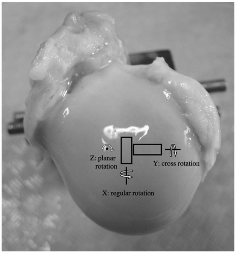
Fig 1.

The orientations of the tissue blocks on the humeral head and the two rotational axes (regular and cross) of the imaging experiments. B0 is in the normal direction of the cartilage surface, in parallel with the z direction. (The planar rotation was not carried out in this project.)
