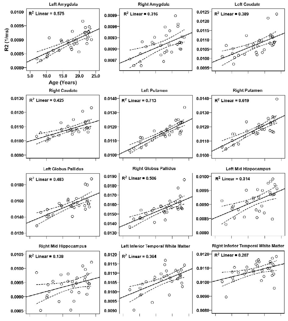
Figure 2.
Brain regions within the rostral brain (telencephalon), including the amygdala, caudate, putamen, globus pallidus, hippocampus, and temporal white matter showing significant correlations between R2 values and age. Solid lines show best fit lines for the data, and dotted lines above and below the best fit line show 95% mean confidence intervals.

