
Figure 1.
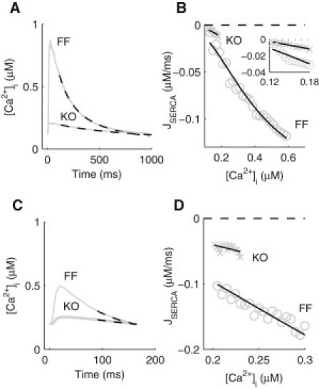
Parameterization of SERCA in the FF and KO models. (A) Average experimentally recorded [Ca2+]i transients, paced at 1 Hz, in isolated ventricular myocytes from the SERCA2 FF and KO mice as indicated. Simulated decay of the [Ca2+]i transients (dashed lines) using the fitted parameter values for SERCA (1 Hz), NCX, and PMCA are superimposed. (B) Ca2+ flux through SERCA (JSERCA) at 1 Hz, plotted as a function of [Ca2+]i, for FF (circles) and KO (crosses) mice. Fitted (JSERCA) are superimposed. Inset: an enlarged view comparing SERCA activity in the KO and FF. (C) Average experimentally recorded [Ca2+]i transients, paced at 6 Hz, in isolated ventricular myocytes from the SERCA2 FF and KO mice as indicated. Simulated decay of the [Ca2+]i transients (dashed lines) using the fitted parameter values for SERCA (6 Hz), NCX, and PMCA are superimposed. (D) Ca2+ flux through SERCA (JSERCA) at 6 Hz, plotted as a function of [Ca2+]i, for FF (circles) and KO (crosses) mice. Fitted (JSERCA) are superimposed.