
Figure 5.
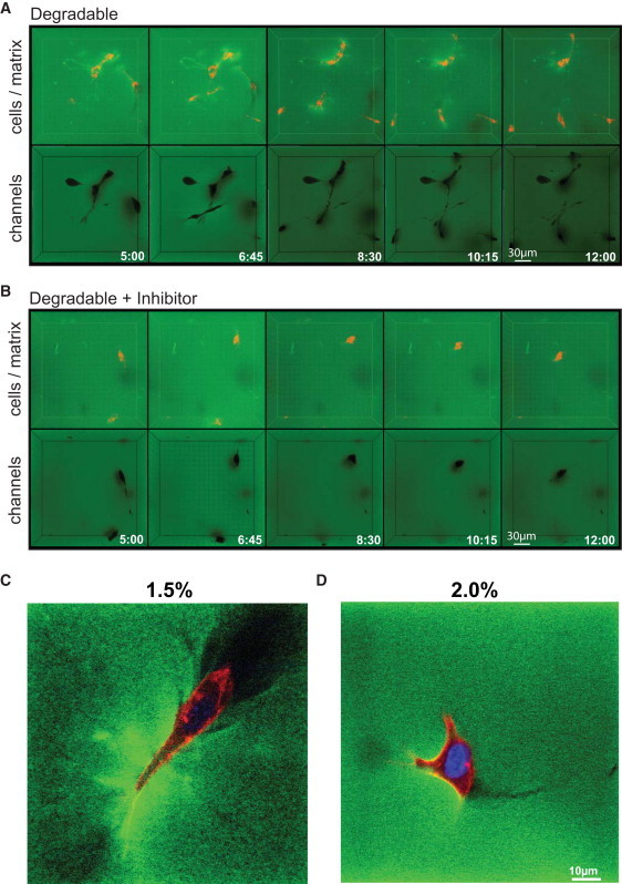
Visualization of cell-induced matrix deformation and macroscopic cavities. (A and B) Migrating PKH26-labeled cells were encapsulated in FITC-conjugated, soft (1.5%) matrices in the absence (A) or presence (B) of GM6001 and followed by four-dimensional time-lapse confocal microscopy for 8 h. In both cases, three-dimensional reconstruction of z stacks over time in maximum intensity projection mode (cells in red, matrix in green) revealed an increase in matrix intensity (green), indicating localized matrix deformation around migrating cells. Three-dimensional reconstruction of stack from the same time points performed in minimum intensity projection mode (matrix in green) revealed the presence of a network of interconnected macroscopic cavities and small cracks indicated the cell migrating paths. (C and D) High-resolution z section of encapsulated cells within soft and intermediate degradable gels after one day in culture (fixed samples).