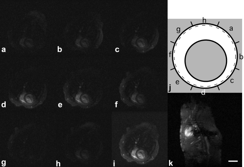
FIG. 1.

a–h: Fully encoded axial in vivo gradient echo images of the individual coil elements. The sum-of-squares reconstruction of (i) axial and (k) sagittal coil images illustrate excellent sensitivity to cover the entire heart in each relevant direction for off-centered positioned mouse. The schematic in panel j depicts the location of the individual coil elements (shown in Fig. 1a–h) relative to the mouse (indicated by the grey circle); the dashed black line represents the inner tube of the probe head, and the solid black circle illustrates the coil-array. Scale bar: 5 mm.
