Fig. 6.
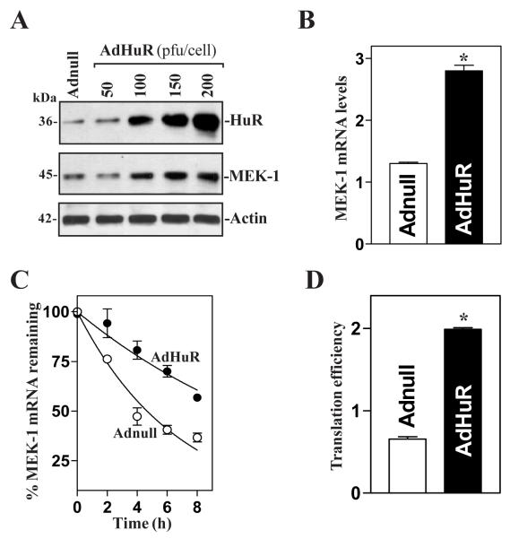
Changes in MEK-1 mRNA stability and translational efficiency after ectopic HuR overexpression. (A) Representative immunoblots of HuR and MEK-1 proteins after ectopic HuR expression. Cells were infected with the recombinant adenoviral vector encoding HuR cDNA (AdHuR, prepared as explained in Materials and Methods) or adenoviral vector lacking HuR cDNA (Adnull) at a multiplicity of infection of 50-200 plaque-forming units (pfu)/cell; the expression of HuR and MEK-1 proteins was analyzed 48 h after the infection. (B) Levels of MEK-1 mRNA as measured by RT-qPCR analysis in cells infected with AdHuR or Adnull at the concentration of 100 pfu/cell for 48 h. Data were normalized to amount of GAPDH mRNA, and values are means ± SE of data from triplicate experiments. * P < 0.05 compared with cells infected with Adnull. (C) Half-life of the MEK-1 mRNA as measured by RT-qPCR analysis by using actinomycin D in cells described in B. Values are the means ± SE from triplicate samples. (D) Changes in MEK-1 translation efficiency as measured by using pGL3-Luc-MEK1ARE reporter assays in cells that were processed as described in B. Twenty-four h after cells were transfected with the pGL3-Luc-MEK1ARE or pGL3-Luc (negative control), the levels of luciferase activity were examined and normalized to the mRNA levels to obtain translation efficiencies. Values were expressed as means ± SE of data from 3 separate experiments. * p < 0.05 compared with cells transfected with Adnull.

