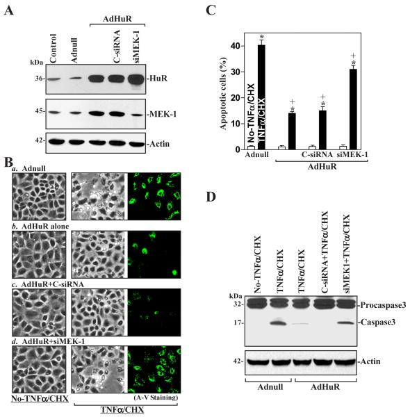
Fig. 8.
Effect of MEK-1 silencing on apoptotic sensitivity in cells overexpressing HuR. (A) Representative HuR and MEK-1 immunoblots. Cells were transfected with either siMEK-1 or C-siRNAfor 24 h and then infected with AdHuR or Adnull (100 pfu/cell). HuR and MEK-1 protein levels were examined by Western blot analysis 24 h after infection, and equal loading was monitored by β-actin immunoblotting. (B) TNFα/CHX-induced apoptosis in cells described A: a) cells infected with Adnull; b) cells infected with AdHuR; c) cells infected with AdHuR and transfected with C-siRNA; d) cells infected with AdHuR and transfected with siMEK-1. Apoptosis was measured by morphological analysis (middle) and ApoAlert annexin-V (A-V) staining (right) 4 h after treatment with TNFα/CHX. Original magnification, ×150. (C) Percentage of apoptotic cells as described in B. Values are means ± SE of data from six samples. * p < 0.05 compared with groups that were not treated with TNFα/CHX. + p < 0.05 compared with cells infected with Adnull and then treated with TNFα/CHX. (D) Representative immunoblots for procaspase-3 and caspase-3 in cells that were processed as described in B. Whole-cell lysates were harvested 4 h after treatment with TNFα/CHX, and the levels of procaspase-3 and caspase-3 proteins were examined by Western blot analysis. Three experiments were performed that showed similar results.

