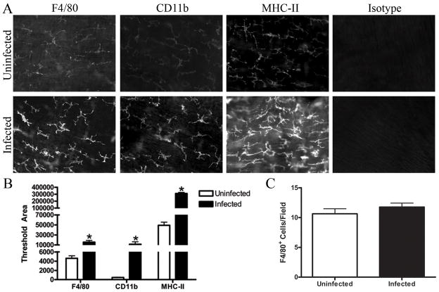
Figure 1. Helicobacter infection increases F4/80, CD11b, and MHC-II expression by intestinal muscularis macrophages.
Whole mounts of intestinal muscle from uninfected or infected mice were stained with F4/80, CD11b, MHC-II, or appropriate isotype control. Representative sections are shown (A) and relative fluorescence of the photomicrographs from uninfected (□) or infected (■) mice is quantified (B). The average number of F4/80 positive cells per field (200x) was calculated using photomicrographs of muscle from uninfected (□) or infected (■) mice (C). Photomicrographs (200X) are representative of 3–4 experiments with 4–6 representative photos analyzed per treatment. *p<0.05 infected compared to uninfected.

