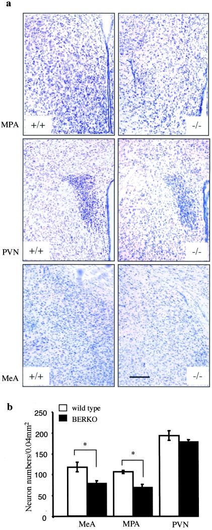
Figure 2.
(a) Coronal sections (30 μm, Nissl stain) of MPA (Top), PVN (Middle), and MeA (Bottom) show histology comparison of 2-yr-old wild-type (+/+) and BERKO (−/−) male mice. Note the decreased number of neurons in these nuclei in BERKO mice. (Scale bar = 170 μm.) (b) Mean number of neurons in MPA, PVN, and MeA (n = 3; error bar, SD; **, P < 0.05, Student's t test).

