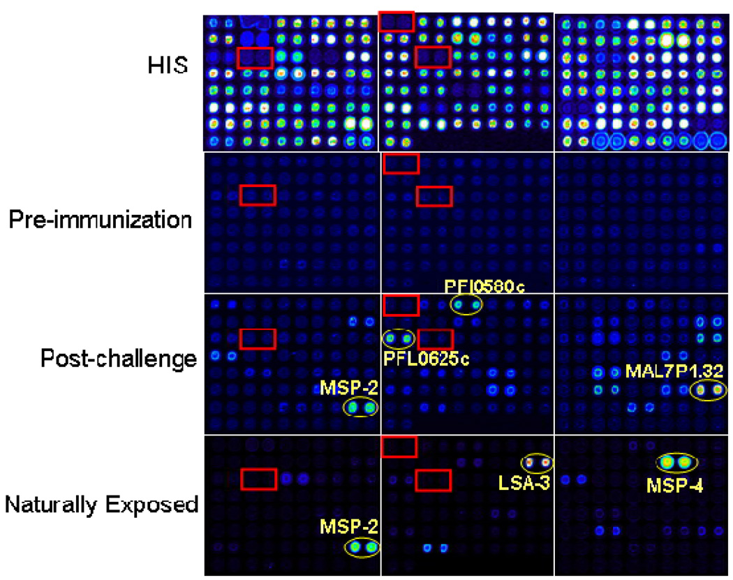
Figure 1. Probed Arrays.
Pf protein microarrays were generated as described in Methods. Three individual arrays displaying a subset of targets evaluated in this study were probed with sera as described below, followed by biotinylated secondary and streptavidin PBXL-3, read in a ScanArray 4000 confocal laser scanner, and the signal intensities quantified. Arrays were probed with (A) anti-polyhistidine mouse monoclonal antibody (B) sera from malaria-naïve individuals (pre-immunization specimens from volunteers subsequently immunized with radiation attenuated Pf sporozoites) (C) sera from irradiated sporozoite immunized and infected volunteers post-challenge, or (D) sera from adults naturally exposed to malaria in Africa. Control reactions that lacked vector template (boxes) and reactions of some well characterized Pf antigens (circles) were also spotted.

