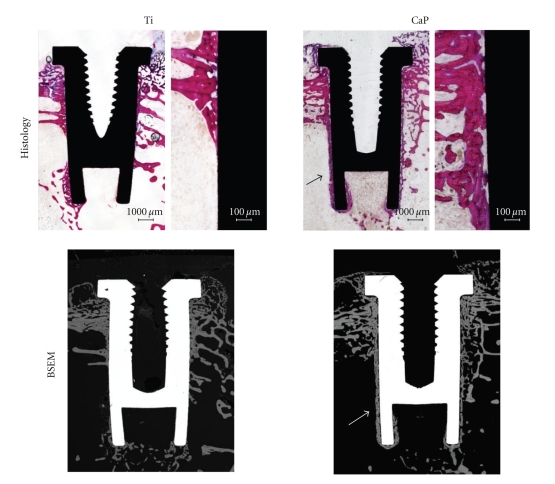
Figure 6.
Micrographs showing the osteointegration of bare titanium- (Ti-) and calcium phosphate- (CaP-) coated implants after implantation in femoral condyles of rabbits for 4 weeks. Note the direct bone apposition on CaP-coated implants (arrows) on both histology (basic fuchsin, toluidine blue staining) and back-scattered electron microscopy (BSEM) images.

