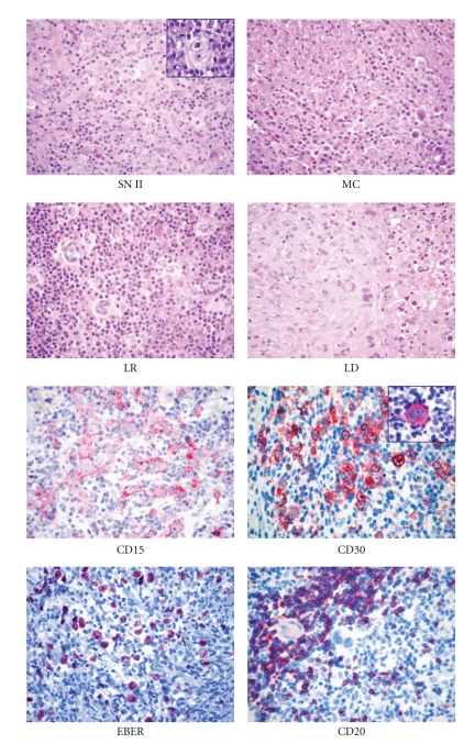
Figure 1.
Histopathological features of classical Hodgkin lymphoma (cHL). At morphology (H&E staining), it is possible to distinguish nodular sclerosis (NS)*, mixed cellularity (MC), lymphocyte rich (LR), and lymphocyte-depleted (LD) subtypes. At immunophenotyping, cHL is typically CD15+, CD30+∗, possibly EBER+, and CD20-/+ (Olympus BX41 microscope, Olympus CAMEDIA C-7070 camera, magnification ×400, colours balanced after acquisition with Adobe Photoshop). *Note the typical Reed-Sternberg cells in the insets.

