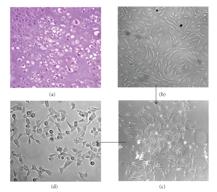
Figure 2.
Generation of a primary cell culture from a chondrosarcoma tissue. (a) Haematoxylin/eosin stained section of a human primary grade 2, focally grade 3 chondrosarcoma of sternum (patient no. 6). (b–d) A representative sample from the tumor excision was subjected to enzymatic digestion. The isolated cells, recovered by centrifugation at 1500 rpm, were cultured in DMEM 10% FBS until to the third passage. At this passage, cell population showed an evident cellular heterogeneity (b). Subcloning of the isolated cell clusters (c) and amplification for six further passages resulted in an adherent, homogeneous cell population characterized by small, chondrosarcoma-like cells (d). Images were captured by an inverted microscope connected to a video camera. Original magnification: x200 (a and c), x400 (b and d).

