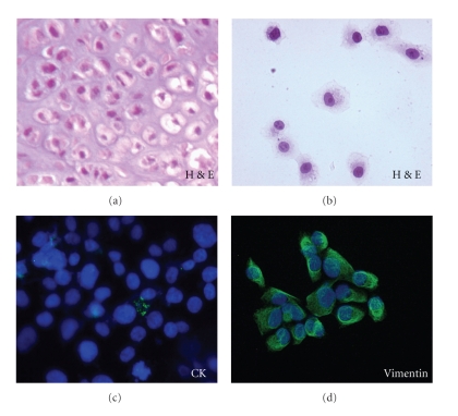
Figure 3.
Immunophenotyping of chondrosarcoma cells. (a, b) haematoxilin/eosin stained chondrosarcoma cells resemble in shape and size those of chondrosarcoma tissue section. (c, d) chondrosarcoma cells grown on glass slides to semiconfluence were stained with anti-cytokeratin (CK) or anti-vimentin mAbs and with Alexa 488-conjugated F(ab')2 fragment of rabbit anti-mouse IgG (green). Nuclei were stained blue with DAPI. Original magnification: x400.

