Figure 1.

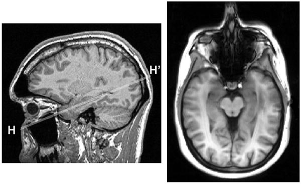
Slice positioning for hippocampal ASL. Left: longitudinal axis H-H’ of the right hippocampus is selected on a sagittal MPRAGE view. Right: resulting ASL axial imaging plane.

Slice positioning for hippocampal ASL. Left: longitudinal axis H-H’ of the right hippocampus is selected on a sagittal MPRAGE view. Right: resulting ASL axial imaging plane.