Figure 3.

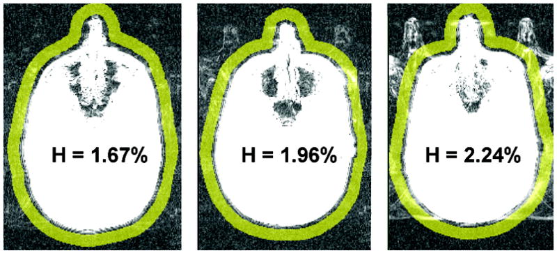
Fringe area (in yellow) used to assess the head motion. Shown are three exams, representing different magnitudes of the head motion index H. H is normalized to the signal in the white matter.

Fringe area (in yellow) used to assess the head motion. Shown are three exams, representing different magnitudes of the head motion index H. H is normalized to the signal in the white matter.